Ultrasound scan: types, preparation and what it can show
An ultrasound scan is one of the most widely used imaging tests – quick, painless and free of X-ray radiation. Doctors use it to look at the abdominal organs, thyroid, breasts, blood vessels, joints and pregnancy. This guide explains the main types of scan, how to prepare and how to make sense of the report.
What is an ultrasound scan?
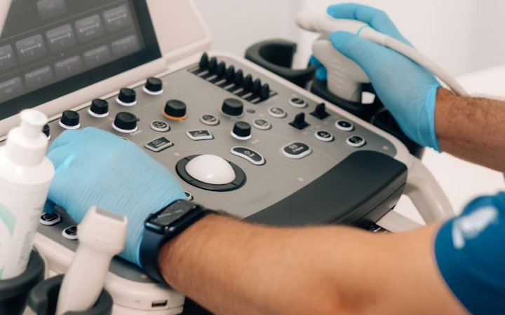
Ultrasound imaging uses sound waves at frequencies too high for the human ear. A hand-held probe (transducer) sends the waves into the body and picks up the echoes that bounce back from tissues. A computer turns these echoes into a live picture, so the doctor can see organs as they move and blood as it flows.
Because there is no ionising radiation, scans can be repeated as often as needed, which is why ultrasound is the first choice in pregnancy and for children. A water-based gel is applied to the skin to remove air between the probe and the body. For some scans the probe is placed inside the body – vaginally, rectally or through the oesophagus for a transoesophageal heart scan.
Ultrasound is one of many tests covered in our diagnostics section. It is often the first scan a doctor orders, before deciding whether MRI or CT is needed.
Types of ultrasound scan
The probe and technique depend on the part of the body and the question being asked:
- Abdominal ultrasound – liver, gallbladder, bile ducts, pancreas, spleen, kidneys and the abdominal aorta. It is commonly used for abdominal pain, gallstones and fatty liver disease.
- Pelvic ultrasound – womb, ovaries, bladder and prostate. In women it can be done through the tummy or transvaginally, for example to assess fibroids.
- Thyroid ultrasound – nodules, size and texture; a suspicious nodule can be sampled with a fine-needle biopsy under ultrasound guidance (see thyroid cancer).
- Breast ultrasound – complements mammography, especially in dense breasts and younger women, and guides a breast biopsy.
- Doppler (vascular) ultrasound – carotid arteries in the neck, linked to stroke risk; leg veins when a clot is suspected or with varicose veins; and leg arteries.
- Heart ultrasound – a separate test described in echocardiography.
- Musculoskeletal ultrasound – tendons, muscles, bursae, shoulder and knee.
- Pregnancy scans – dating, anomaly and growth scans.
How to prepare
Preparation depends on the scan, and the clinic will normally send instructions:
- Abdominal scan: you are usually asked not to eat for about 6–8 hours, so the gallbladder is full and there is less gas in the bowel. Water is usually allowed.
- Pelvic scan through the tummy: you need a full bladder – typically drinking about a litre of water an hour beforehand and not emptying your bladder.
- Transvaginal scan: your bladder usually needs to be empty.
- Thyroid, breast, vascular and joint scans: usually no special preparation.
Bring earlier reports and images if you have them – comparing with a previous scan is often as useful as the new scan itself. Wear clothes that make the area easy to uncover.
What happens during the scan
You lie on a couch, gel is applied and the sonographer or doctor moves the probe over your skin. You may be asked to take a deep breath and hold it, or to roll onto one side. Most scans take 10–30 minutes and are painless, although pressure from the probe can be uncomfortable over a tender area or a full bladder.
For internal scans the probe is slim and covered with a protective sheath; there may be mild discomfort. A contrast-enhanced ultrasound, used in specialist centres for example to characterise liver lesions, needs a small cannula in a vein.
You can go back to normal activities straight away. Findings are often discussed during the scan, and a written report is usually available the same day.
What ultrasound can and cannot show
Ultrasound is very good at telling fluid from solid tissue – a simple cyst from a solid lump – and at assessing the gallbladder, kidneys, thyroid and blood vessels. It is also the standard way to guide a needle for a biopsy or drainage.
It has limits too:
- sound waves do not pass well through air or bone, so the bowel, lungs and adult brain are poorly seen;
- images are less clear in people with a higher body weight;
- results depend heavily on the operator and the quality of the machine;
- a normal scan does not rule out every condition – small pancreatic tumours, for example, are often missed.
| Test | Radiation | Best suited to |
|---|---|---|
| Ultrasound | None | Abdominal organs, thyroid, breast, blood vessels, pregnancy |
| CT | X-rays | Lungs, bones, rapid abdominal assessment, cancer staging |
| MRI | None (magnetic field) | Brain, spine, joints, pelvis, liver |
Understanding your report
The report describes organ size, texture (“echogenicity”) and any findings. A few common phrases:
- “Echogenic” or “bright” liver – brighter than normal, often a sign of fatty liver.
- “Anechoic lesion” – usually fluid, such as a simple cyst, which is most often harmless.
- “Hypoechoic solid nodule” – solid tissue; depending on its features it may need monitoring or a biopsy.
- Scoring systems – BI-RADS for the breast and TI-RADS-type systems for thyroid nodules help decide whether a biopsy is needed.
A report on its own is not a diagnosis. The doctor who requested the scan interprets it alongside your symptoms and other results. If findings are worrying or unclear, a second opinion from a specialist is a sensible step.
Ultrasound in health check-ups and abroad
Ultrasound features in most health check-up packages: abdominal scan, thyroid, breast and pelvis in women, prostate in men. In several countries older men who have smoked are offered a one-off ultrasound to screen for an abdominal aortic aneurysm.
Tests are best chosen according to age, sex and risk factors rather than “everything for everyone”: unnecessary scans sometimes find harmless incidental findings that lead to anxiety and further tests. Read more in our guide to a sensible health check-up.
Some patients from Bulgaria and elsewhere in Europe combine a full assessment with a trip to Istanbul – see check-up in Turkey – where large hospitals usually perform imaging, blood tests and specialist consultation on the same day. New City Clinic arranges such check-ups with a coordinator and interpreter. Whatever you choose, ask for a written report in English so that your own doctor can use it.
When to see a doctor
See your GP, who will decide whether you need a scan and which one, if you have:
- abdominal pain, especially under the right ribs after eating;
- a lump in the neck, breast, groin or elsewhere;
- changes in passing urine, blood in the urine or loin pain;
- a swollen, painful leg;
- heavy or irregular periods or pelvic pain.
Seek emergency care (call 112) for sudden severe abdominal pain, a swollen leg together with breathlessness or chest pain, or bleeding in pregnancy.
Frequently asked questions
Is ultrasound harmful?
Diagnostic ultrasound does not use ionising radiation and has no proven harmful effects when used for a medical reason. That is why it is the main imaging test in pregnancy and in children. It is still best done only when there is a clinical reason.
Do I need to fast before an ultrasound?
For an abdominal scan, usually yes – around 6–8 hours without food, though water is normally allowed. Thyroid, breast, joint and most vascular scans need no preparation. Always follow the clinic’s instructions.
What is the difference between ultrasound and Doppler?
A standard scan shows the structure of organs. Doppler ultrasound measures blood flow and shows whether a vessel is narrowed or blocked, for example a clot in a leg vein or narrowing of a carotid artery.
Can ultrasound detect cancer?
Ultrasound can show lumps that look suspicious in organs such as the liver, kidneys, thyroid or breast. A definite diagnosis, however, needs a biopsy and laboratory analysis, often with further imaging.
How soon will I get the results?
The person scanning often discusses what they see during the test, and a written report is usually ready the same day. If another specialist requested the scan, they should interpret it in the light of your symptoms.
Sources
This article is for education and does not replace consultation, diagnosis or treatment by a doctor. In an emergency call 112.