Echocardiogram: what it shows, how it is done and what the results mean
An echocardiogram is an ultrasound scan of the heart that shows, in real time, how the chambers pump, how the valves open and close and whether there is fluid around the heart. It is painless, uses no radiation and usually takes 20 to 45 minutes.
What is an echocardiogram?
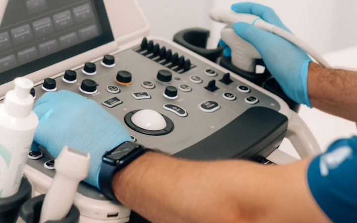
An echocardiogram, often shortened to "echo", is a type of ultrasound scan. A probe sends high-frequency sound waves into the chest; they bounce back from the structures of the heart and the machine turns them into a moving image. The cardiologist can watch the heart relax and contract, see the valves working and follow the blood as it flows.
Doppler measurements add information on the speed and direction of blood flow. This shows whether a valve is narrowed (stenosis) or leaking (regurgitation) and allows an estimate of the pressure in the lungs' circulation.
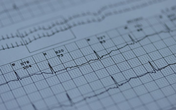
Echo is one of the most widely used tests in cardiology and is central to the assessment of most heart conditions. It complements, but does not replace, an electrocardiogram (ECG), which records the heart's electrical activity.
Why might you need an echo?
Your doctor may request an echocardiogram in many situations, for example:
- breathlessness, swollen ankles or unusual tiredness – to look for signs of heart failure;
- a heart murmur heard with a stethoscope – to assess heart valve disease;
- chest pain or a previous heart attack – to see whether part of the heart muscle is not contracting normally;
- heart rhythm problems such as atrial fibrillation – to assess the upper chambers and look for an underlying cause;
- long-standing high blood pressure – to check whether the wall of the left ventricle has thickened;
- a suspected congenital heart defect, endocarditis (infection of the valves), fluid around the heart or cardiomyopathy;
- before and after heart surgery, and for monitoring during some cancer treatments that can affect the heart.
Types of echocardiogram
| Type | How it is done | Typical use |
|---|---|---|
| Transthoracic (TTE) | Probe moved over the chest with gel | The standard test for most questions |
| Transoesophageal (TOE/TEE) | A thin probe is passed through the mouth into the oesophagus, with a throat spray and usually light sedation | Looking for clots in the atria, endocarditis, detailed views of valves and valve replacements, before certain procedures |
| Stress echo | Images recorded at rest and during exercise (treadmill or bike) or after a medicine that makes the heart work harder | Looking for reduced blood supply in angina, assessing some valve problems |
| Contrast echo | An ultrasound contrast agent (tiny bubbles) is injected into a vein | When images are hard to obtain, or to detect a hole between the atria |
| 3D echo | A special probe creates a three-dimensional image | Detailed valve assessment and surgical planning |
Echocardiograms in young children and during pregnancy (fetal echo) are performed by specialists experienced in those areas.
What happens during the test and how to prepare
A standard transthoracic echo needs no special preparation. You can eat, drink and take your medicines as usual. You will be asked to undress to the waist and lie on your left side. ECG stickers are placed on your chest and gel is applied to the skin. A cardiologist or trained cardiac physiologist moves the probe to different positions and may ask you to hold your breath briefly. It does not hurt, although you may feel some pressure.
For a transoesophageal echo you will usually need to stop eating and drinking for several hours beforehand – the hospital will give precise instructions. Your throat is numbed with a spray and light sedation is often given. Afterwards you should not drive for the rest of the day, and you should not eat until feeling has returned to your throat.
For a stress echo, wear comfortable clothes and shoes. Your doctor will tell you whether any medicines, such as beta-blockers, should be paused beforehand – never stop them on your own.
Understanding your echo report
An echo report is full of numbers and terms. The most important ones are:
- Ejection fraction (EF) – the proportion of blood in the left ventricle pumped out with each beat. Values of roughly 55–70% are usually considered normal; lower values may indicate reduced pumping function. Your doctor interprets the figure in context.
- Chamber sizes and wall thickness – enlarged chambers or thickened walls can point to high blood pressure, valve disease or cardiomyopathy.
- Wall motion – an area that does not contract normally may be the result of a previous heart attack.
- Valves – whether there is stenosis or regurgitation and how severe it is (mild, moderate, severe).
- Diastolic function – how well the heart relaxes and fills.
- Pericardium – whether there is fluid around the heart.
- Pulmonary pressure – an estimate of pressure in the lung arteries.
Trivial or mild leakage of a valve is a common finding and often of no significance. Avoid drawing conclusions from the report alone – go through it with your cardiologist.
Benefits and limitations
Echocardiography is safe, quick, widely available and can even be done at the bedside of a very ill patient. It has no known harmful effects and is suitable during pregnancy.
It has limitations too. Obesity, lung disease or chest wall shape can make images less clear, and quality depends on the operator's experience. Echo does not show the coronary arteries directly – if narrowing is suspected, a stress test, CT coronary angiography or invasive coronary angiography may be needed. Sometimes a cardiac MRI scan (see MRI and CT) gives a more detailed picture of the heart muscle.
Transoesophageal echo carries small risks related to the probe and sedation, such as a sore throat and, rarely, injury to the oesophagus. These are explained before you consent.
When to see a doctor
See your GP, who can refer you to a cardiologist, if you become breathless with everyday activity, notice swollen ankles, palpitations, unexplained tiredness, dizziness or near-fainting episodes. If you already have a heart condition, keep to your scheduled check-ups and repeat scans.
Call 112 (or your local emergency number) for severe or prolonged chest pain, sudden severe breathlessness, collapse or signs of a stroke – do not wait for a planned test.
Echocardiography and cardiac assessment abroad / in Türkiye
Echocardiograms are widely available across Europe. Some patients from Bulgaria and elsewhere nevertheless look for a second opinion or a complete cardiac work-up abroad, particularly before heart surgery or when 3D or transoesophageal echo is needed. Some choose large accredited hospital groups in Istanbul, where echo is often part of a broader health check-up or cardiology package.
If you consider this, check the hospital's accreditation and the cardiologist's experience, make sure you will receive a written report and copies of the images, and agree how the results will be reviewed with your cardiologist at home. Organisers such as New City Clinic in Istanbul coordinate hospital, doctor, hotel, transfers and interpreter, while all medical decisions remain with the treating team. Our diagnostics guide explains other common tests.
Frequently asked questions
Does an echocardiogram hurt?
A standard transthoracic echo does not hurt – you will only feel the gel and some pressure from the probe. A transoesophageal echo can be uncomfortable, but the throat is numbed and light sedation is usually given.
How long does an echo take?
Usually 20 to 45 minutes. Transoesophageal and stress echo take longer once preparation and recovery time are included.
Do I need to fast before an echo?
Not for a transthoracic echo. For a transoesophageal echo you will normally need to avoid food and drink for several hours; follow the hospital's instructions.
Is an echocardiogram safe?
Yes. It uses ultrasound rather than X-rays and can be repeated as often as needed. It is also safe in pregnancy.
What does a low ejection fraction mean?
A low ejection fraction means the left ventricle is pumping out less blood than normal with each beat. It can be a sign of heart failure, but its significance is judged by a cardiologist together with your symptoms and other tests.
Sources
This article is for education and does not replace consultation, diagnosis or treatment by a doctor. In an emergency call 112.